Left Ventricle Ejection Fraction (LVEF)
The Left Ventricle Ejection Fraction (LVEF) function is used to estimate the volume of blood pumped out by the left ventricle of the heart.
The LVEF measurement is based on estimation of the volume of left ventricle at two different states: End Diastole (when the left ventricle is at its maximum volume) and End Systole (when the left ventricle is at its minimum volume). The volume of the left ventricle is estimated using the Simpson method on cardiac ultrasound images in Apical 4 Chamber (A4C) and Apical 2 Chamber (A2C) views.
Note
It is more convenient to perform this measurement in 1x1 view. Switch to 1x1 view if the current view is different.
- Begin the measurement by loading an ultrasound series with an Apical 4 Chamber view of the heart.
- If Cine is playing, stop it by selecting the Cine Stop button on the toolbar.
Finding diastole
Find the image of end diastole inside the series by using the ECG signal as an aid, or visually locating the image with maximum relaxation of the left ventricle.
Finding systole
With the current series still selected, locate and open the image of end systole by using the ECG signal as an aid, or visually locating the image with maximum contraction of the left ventricle.
Measuring the volume using the Simpson method
- From the menu bar, select Annotation | Cardiology | Simpson Volume.
- To start marking the area of the left ventricle, click in the corner at one end of the mitral valve annulus, and drag the mouse around to the other end of the annulus, encapsulating the entire chamber. When the mouse is released, the Simpson Volume Properties dialog box is displayed.
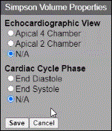
- Under Echocardiographic View, select the type of the view in this image.
- Under Cardiac Cycle Phase, select the phase to which this cycle corresponds.
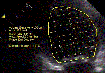
- Click Save. An information box displays on the image and contains the measurement details including volume, area, major axis, view, and phase. If an end systole measurement has not been performed, the Ejection Fraction information is not shown.
Notes
The Ejection Fraction value will be displayed only after volumes for both End Diastole and End Systole phases are measured. The Biplane label on volume measurement means that the measurement uses biplane Simpson method, which requires performing the measurement on both Apical 4 Chamber and Apical 2 Chamber views as will be explained further below.
You can adjust the position and direction of the major axis of the ventricle by moving the apex point along the ventricle contour. Specifically, you can:
- Adjust end points of the contour by moving them slightly
- Adjust direction of the major axis by moving the apex point along the ventricle contour
- Adjust part of the contour by redrawing only this portion, beginning at some position on the contour
- As you make the adjustments, measurement values will be updated
Selecting the end systole
- With the current series still selected, locate and open the image of end systole by using the ECG signal as an aid, or visually locating the image with maximum contraction of the left ventricle.
- Repeat steps 1-6 outlined in the previous section.
The measurements made are automatically saved. In addition, after the Simpson Volumes are measured for end diastole and end systole, the ejection fraction value is computed and displayed on each of the measurements.
Improving precision using biplane Simpson volume
If the study contains a series of Apical 2 Chamber views, additional measurements can be performed on this view to improve precision of the left ventricle volume and ejection fraction.
- Select the series with the Apical 2 Chamber view.
- Measure the Simpson Volumes at the end diastole and end systole points, as done for the Apical 4 Chamber view.
- From the menu bar, select Annotation | Cardiology | Simpson Volume.
- In the Simpson Volume Properties dialog box, select Apical 2 Chamber.
- Select the appropriate Cardiac Cycle Phase.
The following occurs:
- The values of the volumes are computed using biplane Simpson method.
- The value of ejection fraction will be updated.
- The values are updated on the measurements of the Apical 4 Chamber view as well.
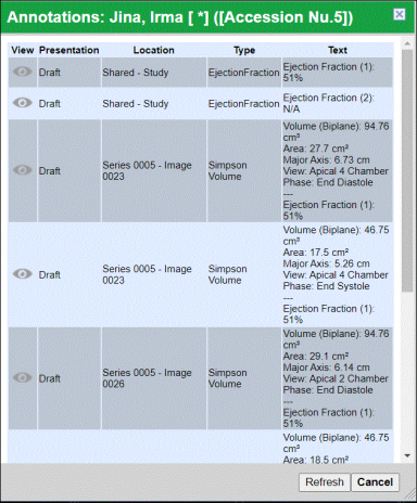
Viewing all volumes used for ejection fraction calculation
There are two methods for viewing the calculation volumes:
Method 1:
Right-click on any of the Simpson volumes and select Show Related volumes.
Method 2:
- Right-click on any image in the study.
- Select View Study Annotations.
- Select the ejection fraction measurement.
- Click the View icon.
Replacing one volume with another
It is possible to recompute the existing ejection fraction measurement by again measuring a volume with particular cardiac view and phase. This can be useful if a better image is found, and would like to use that image instead.
- Measure the Simpson Volume on a new image as described under Measuring the volume using the Simpson method. The Warning dialog box is displayed.
-
Select Overwrite to replace a previously measured Simpson Volume with the new one. The ejection fraction value will be recomputed, and the values will be updated on all other Simpson Volume measurements in the study.
Performing another ejection fraction measurement in the same study
It is possible to measure ejection fraction more than once using a different set of images.
- Select an image with A4C view.
- Start with measuring the Simpson Volume at end diastole point (as described previously). The Warning dialog box is displayed.
- Select New to add the new volume measurement and start a new ejection fraction measurement. The previous ejection fraction measurement remains unchanged and becomes immutable and cannot be updated.
- Continue measuring additional Simpson Volumes (as described above) to complete this ejection fraction measurement.
- To view all measured Simpson Volumes and ejection fractions, right-click on any image.
- From the menu, select View study annotations. The Annotations dialog box is displayed and contains information about an annotation, and includes a View icon, which can be selected to open the image with selected measurement.